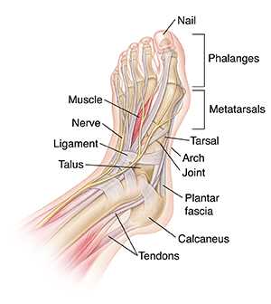
Understanding the Foot Anatomy and Its Key Body Parts
Exploring the structure of the lower extremity is essential for understanding how it supports movement and balance. This complex area is composed of several interconnected regions, each with a distinct function. By examining these areas, one can gain insight into how they contribute to mobility, stability, and overall health.
Key regions in this section offer crucial support and flexibility, enabling various activities from walking to running. These areas work together harmoniously, allowing for a range of motion while providing necessary strength. Understanding their structure can help in identifying potential issues and improving overall function.
The different segments within this region play specific roles in maintaining balance and absorbing impact. Each contributes to the efficiency of movement, and a closer look at their arrangement can reveal much about their importance in daily physical activities.
Anatomy of the Foot: Key Structures
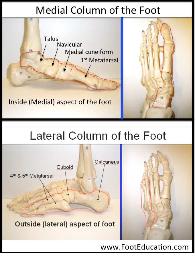
The lower extremity is a complex network of interconnected elements that provide stability, movement, and support. These components work together to ensure proper balance and function in various activities, from standing to walking. Understanding the main features of this region helps to appreciate how essential it is to human mobility.
Main Structural Elements
- Bones: A set of bones forms the foundation of this part of the limb, providing both rigidity and flexibility for movement.
- Muscles: Several muscle groups assist in movements such as flexing, extending, and rotating, playing a vital role in overall strength and control.
- Ligaments: These connective tissues hold the bones together, ensuring stability and preventing excessive movement.
- Tendons: These robust fibers connect muscles to bones, transmitting force that facilitates various motions.
Key Functions
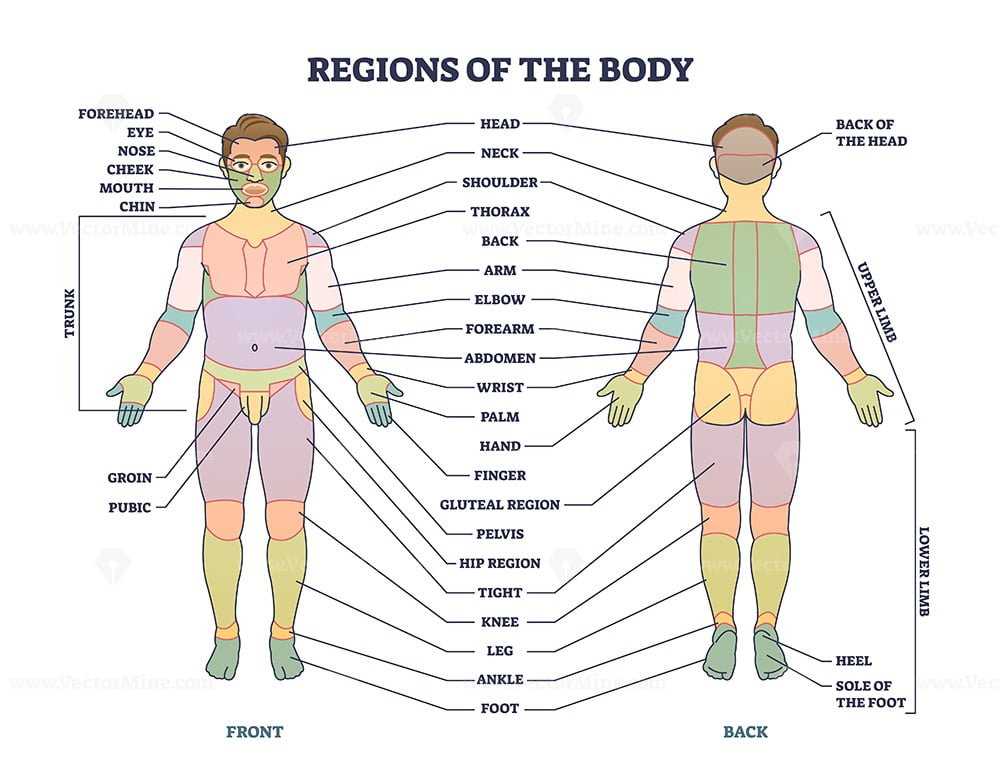
The main functions of this area include maintaining balance, absorbing impact, and providing propulsion during walking or running. These roles are made possible by the coordinated efforts of the bones, muscles, and connective tissues.
Understanding the Arch of the Foot
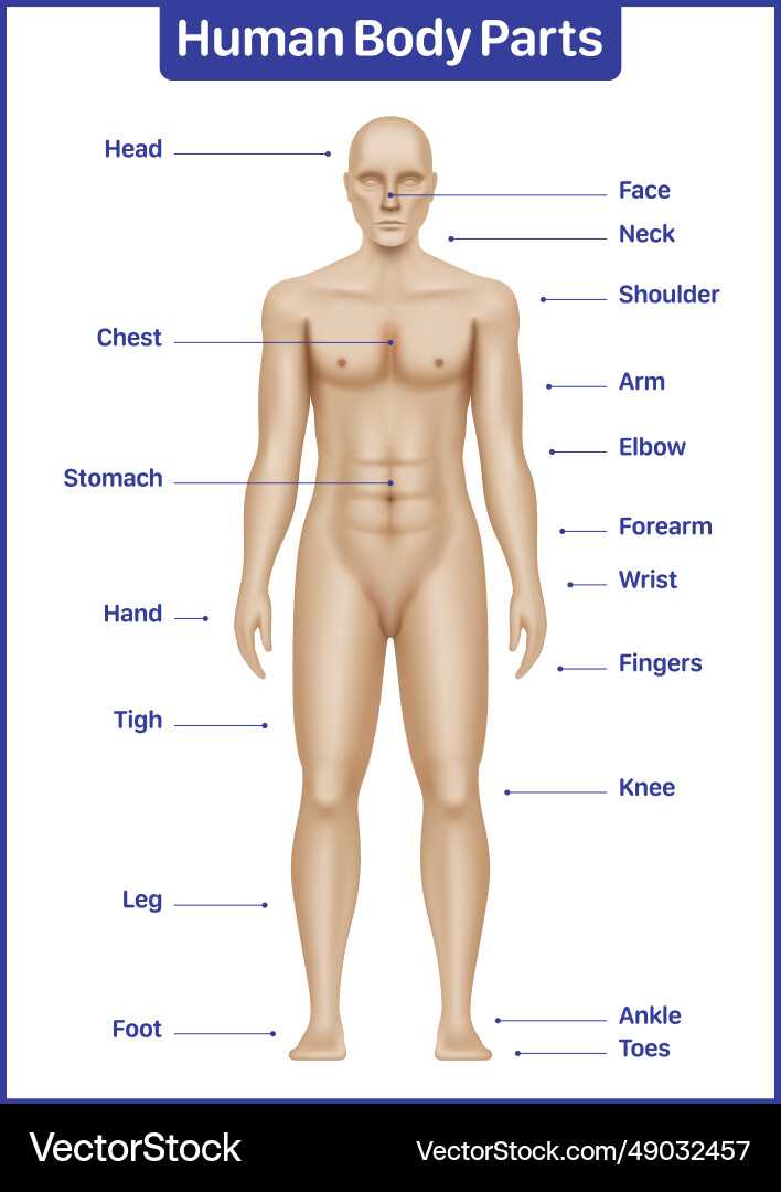
The structure beneath our legs plays a crucial role in movement and balance. It serves as a natural support system that absorbs impact and provides stability during activities like walking or running. The intricate design allows for flexibility while also ensuring strength when carrying weight.
One key aspect of this region is its ability to adjust to different surfaces, maintaining an even distribution of pressure. Without this natural mechanism, movements would be far less efficient, potentially leading to discomfort or strain.
Maintaining the well-being of this area is essential for overall mobility. Simple care practices can ensure that the area remains in optimal condition, reducing the risk of injury or fatigue.
Main Functions of Toes in Movement
Each small extension at the end of our limbs plays a significant role in maintaining balance and aiding locomotion. Their structure and position allow for essential support, helping us navigate different surfaces with stability and control. Without these elements, simple movements would become far more difficult, impacting coordination and fluidity.
Balance and Stability
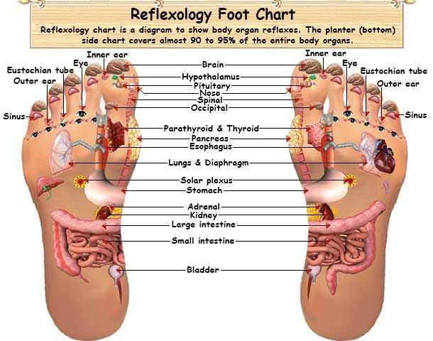
The front extremities help maintain equilibrium, especially during walking or running. They act as an anchor, ensuring proper weight distribution. This allows for smooth transitions from one step to the next, making it easier to stand upright on uneven surfaces.
- Distributes body weight evenly
- Prevents falls and helps regain balance
- Adjusts position to handle different terrains
Propulsion and Movement
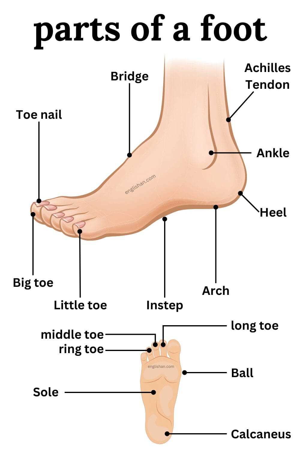
These extensions also serve a key role in pushing off the ground, providing the necessary force to propel forward. By flexing and gripping the surface, they enhance both speed and agility, making movements quicker and more efficient.
- Creates push for forward motion
- Increases speed and momentum
- Improves agility during physical activities
The Role of the Heel in Support
The heel serves as a crucial element in maintaining balance and absorbing impact during movement. It plays a vital role in ensuring stability when standing or walking, working in conjunction with other lower extremities to distribute weight effectively. Understanding its function is key to preventing strain and enhancing overall comfort.
Impact Absorption and Shock Distribution
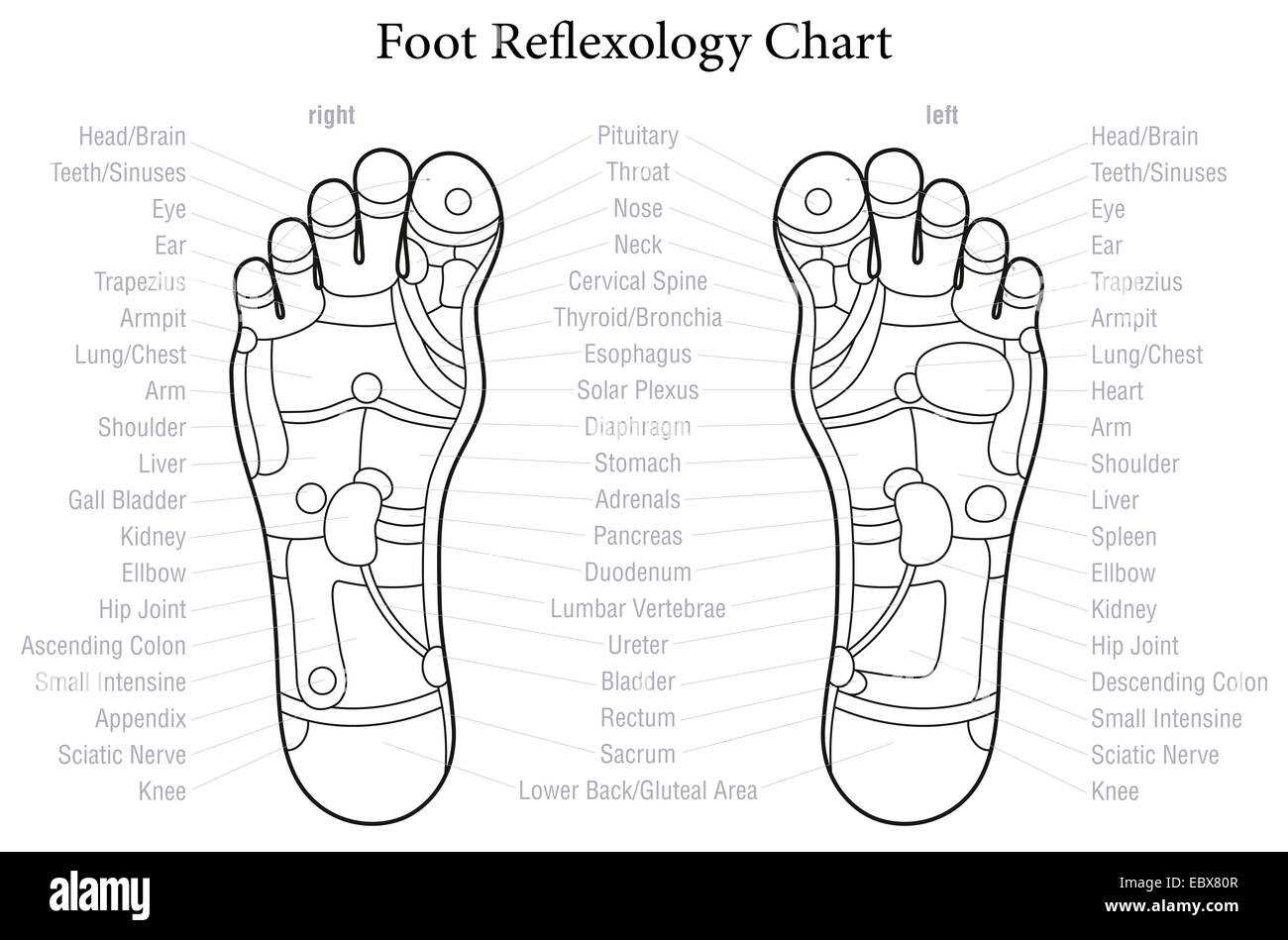
When stepping, the heel is often the first point of contact with the ground. It absorbs much of the force generated by movement, reducing the pressure on other joints and muscles. This helps in minimizing wear and tear over time, ensuring the legs and back remain protected from excessive strain.
Stability and Weight Distribution
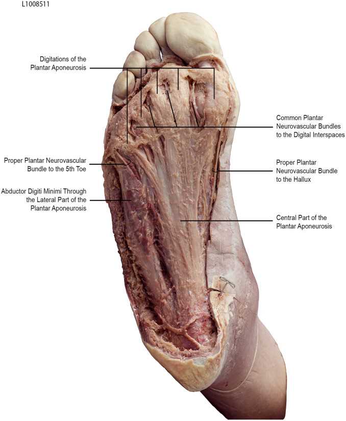
Besides shock absorption, the heel ensures that weight is evenly distributed across the entire support structure. This helps maintain proper posture and allows for smooth transitions between standing, walking, and running, preventing fatigue and improving endurance.
| Function | Benefit | ||||||||||||
|---|---|---|---|---|---|---|---|---|---|---|---|---|---|
| Shock absorption | Reduces strain on joints | ||||||||||||
| Stability | Improves balance | ||||||||||||
| Weight distribution | Enhances posture and movement efficiency |
| Tissue Type | Function | Impact on Movement |
|---|---|---|
| Elastic Connective Tissue | Allows stretching and recoiling | Enhances mobility and range of motion |
| Fibrous Connective Tissue | Provides support and structure | Enables controlled movement while maintaining stability |
| Collagen Fibers | Contribute to strength and resilience | Assist in preventing overextension and injury |
Muscle Groups in the Foot and Their Functions
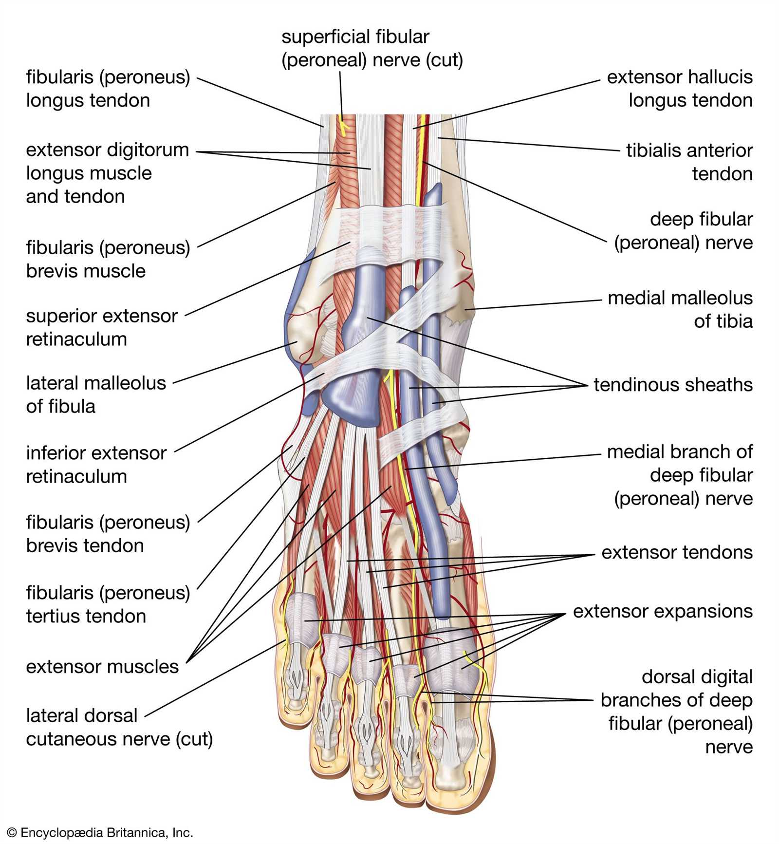
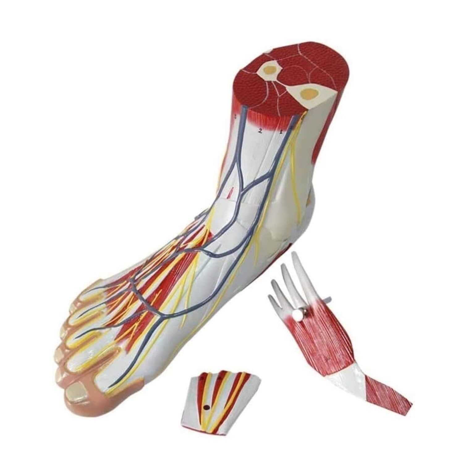
The lower extremity contains various muscle groups that play a crucial role in providing stability and facilitating movement. These muscles are responsible for a range of actions, from basic balance to complex movements during walking, running, and other activities. Understanding the functions of these muscle groups helps in grasping the intricate mechanisms that allow for efficient locomotion and support.
Intrinsic muscles are located within the region, mainly focusing on the toes and arches. They assist with fine motor control, providing support for stability and propulsion. These muscles are key to controlling the position and flexibility of the toes, contributing to a more balanced stance.
Extrinsic muscles, originating from the lower leg, have a broader impact. They are responsible for more significant movements such as dorsiflexion, plantarflexion, and inversion. Their influence extends beyond support, aiding in larger scale motions needed for walking and running, offering the power and mobility required for efficient motion.
Nerve Distribution and Sensation in the Foot
The lower limb is equipped with a complex network of nerve fibers that play a crucial role in sensory perception. These nerves are responsible for transmitting various sensations such as touch, pressure, and pain. This intricate system ensures that the foot responds effectively to external stimuli, maintaining balance and coordination during movement.
Key Nerves and Their Functions
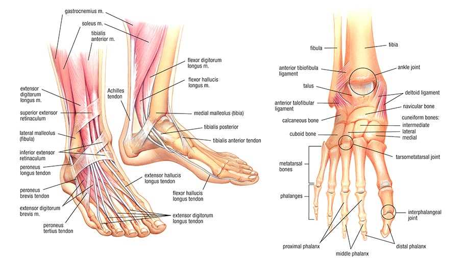
Several nerves contribute to the sensation in this area, each with a distinct role. The tibial nerve primarily governs sensations in the sole, while the peroneal nerve controls the top surface. These two key pathways collaborate to ensure smooth communication between the lower limb and the brain, enabling precise movements and reactions.
Sensory Receptors and Feedback
Sensory receptors within the skin and deeper tissues provide constant feedback to the nervous system. These receptors detect pressure, temperature changes, and painful stimuli. The body can then react to avoid injury or adjust posture, highlighting the vital role of nerve networks in maintaining proper function and stability.
Common Injuries Affecting Foot Anatomy
Various types of damage to the lower extremities can lead to significant discomfort and long-term complications. These issues often arise from overuse, trauma, or improper posture. Some of the most common conditions that affect this area of the body can disrupt mobility and reduce overall quality of life.
Sprains are a frequent injury, usually caused by a sudden twisting or stretching of the ligaments. This often results in pain, swelling, and limited movement. Similarly, fractures of the bones in this region can happen due to impacts or stress over time. These injuries may require immobilization or surgical intervention to ensure proper healing.
Another common issue is tendinitis, which involves inflammation of the tendons due to repetitive stress. This condition typically causes sharp pain and stiffness, making it difficult to perform everyday tasks. Plantar fasciitis, a condition that affects the tissue at the bottom of the foot, also leads to persistent discomfort, especially after long periods of standing or walking.
In some cases, chronic conditions such as arthritis may develop, leading to inflammation and degeneration of the joints, which can cause stiffness and ongoing pain. Proper care, timely treatment, and preventive measures are essential to manage these ailments and maintain mobility.
