Understanding the Key Parts of the Skull Diagram
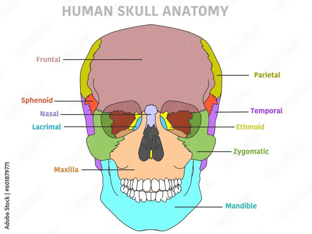
Understanding human cranial structure is essential for various fields, from medicine to anthropology. This intricate framework not only protects vital organs but also provides insight into evolutionary processes and human development.
Within this fascinating realm, one can discover numerous elements, each with distinct functions and characteristics. These components work harmoniously to form a protective casing around the brain, showcasing a remarkable design.
In this exploration, we will delve into different sections, highlighting their significance and interconnections. By grasping this complex architecture, we can appreciate both its functionality and the ultimate beauty of human anatomy.
Understanding the Skull Structure
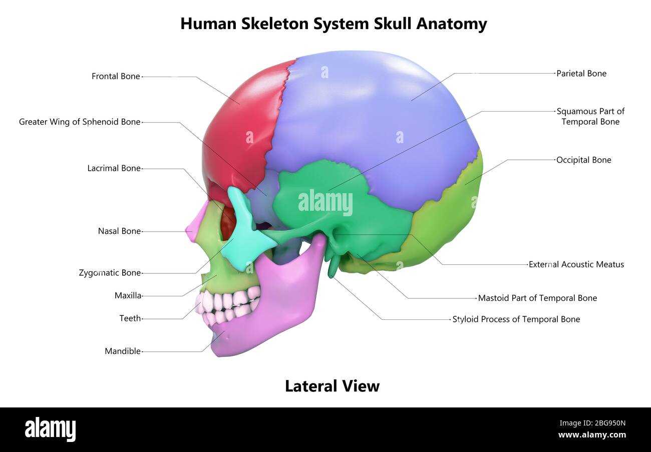
Exploring cranial architecture reveals intricate designs essential for protecting vital organs and supporting various functions. This framework plays a crucial role in shaping facial features and housing sensory organs. A deeper look into its composition helps appreciate its complexity and significance.
Key components can be classified into several categories:
- Protective Elements: These structures create a shield for the brain and provide a sturdy framework.
- Supportive Structures: They maintain the overall shape and alignment, crucial for physical integrity.
- Facial Framework: This includes features that define individual appearance and assist in functions like eating and speaking.
Additionally, understanding the interconnectedness of these components offers insight into both evolutionary development and clinical relevance. Some important structures include:
- Frontal Region
- Parietal Areas
- Temporal Zones
- Occipital Section
By analyzing each segment, one gains a comprehensive view of how these formations interact and contribute to overall functionality, highlighting their importance in both health and disease contexts.
Major Bones of the Human Skull
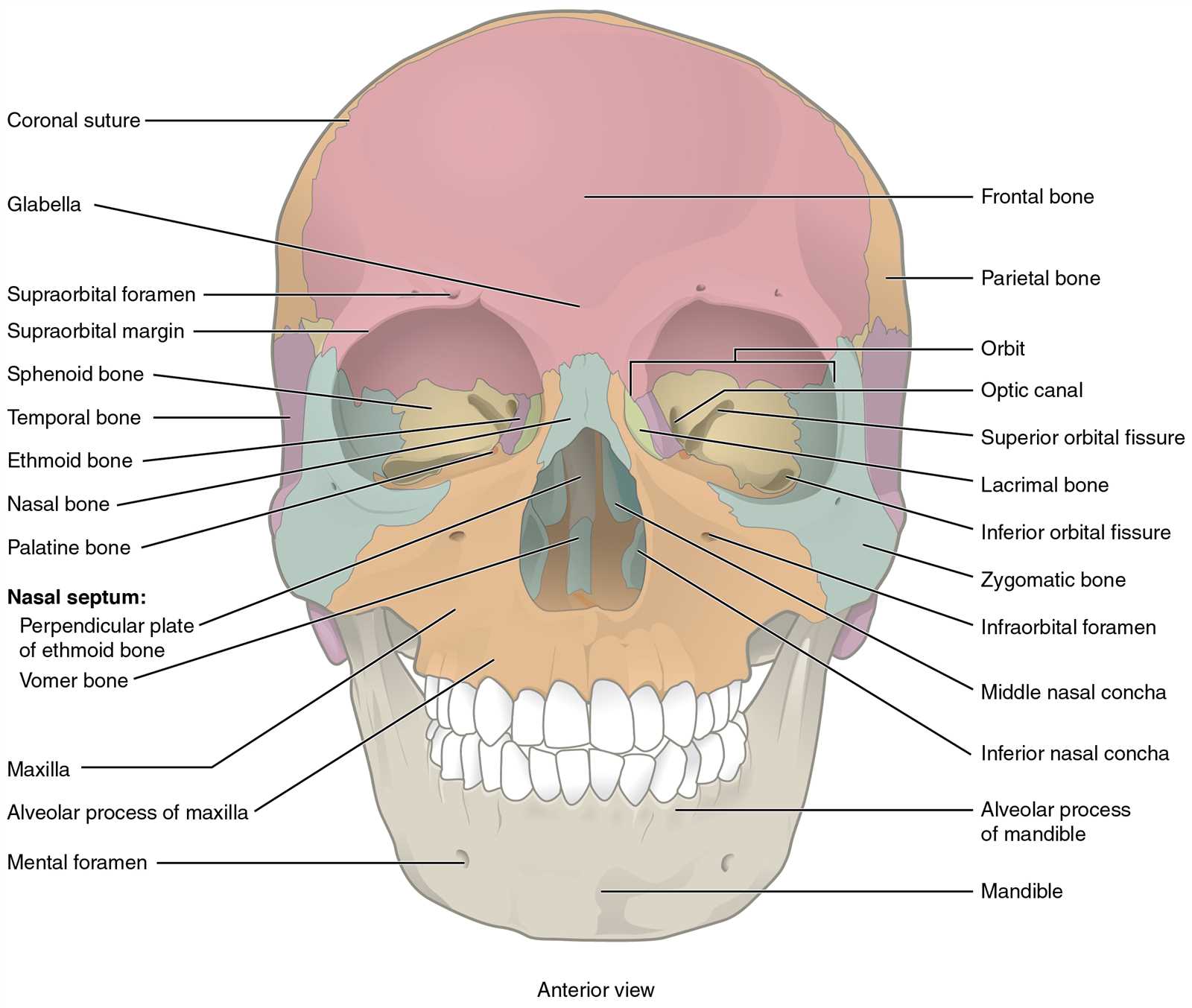
This section explores essential structures that compose the framework of the head. These components play vital roles in protecting delicate organs and supporting facial features.
Key Elements
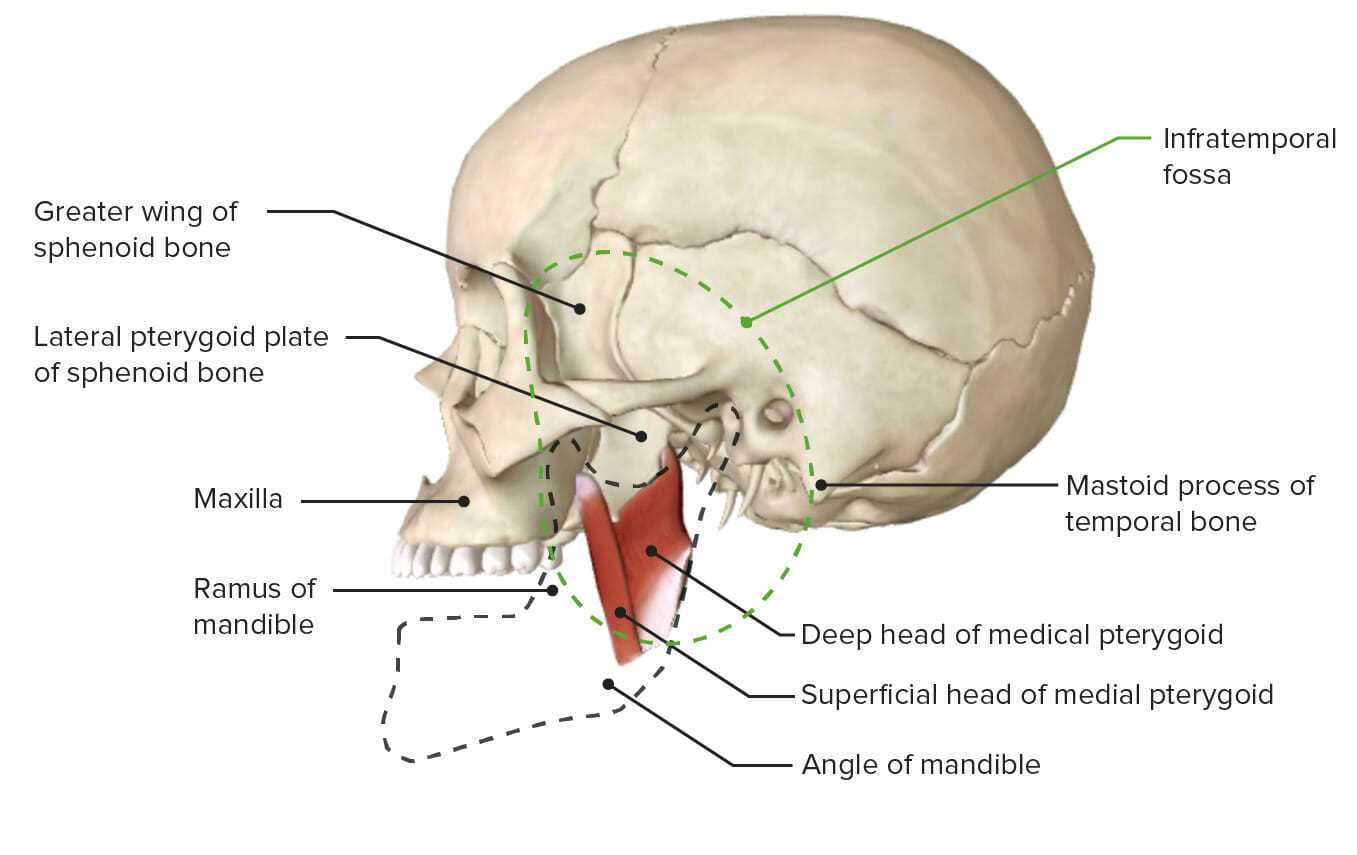
Several primary formations can be identified, each contributing uniquely to overall functionality and form. Understanding their characteristics enhances appreciation for human anatomy.
Overview Table
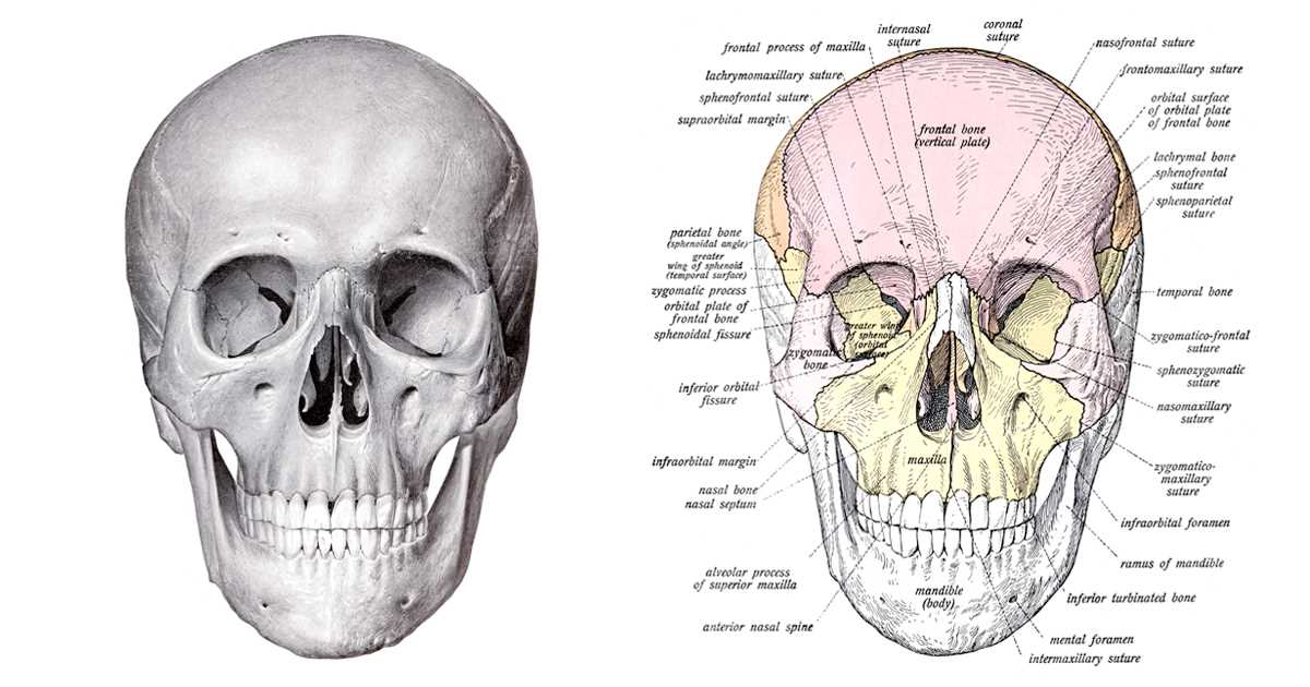
| Bone Name | Location | Function |
|---|---|---|
| Frontal | Forehead area | Protects brain; forms upper facial structure |
| Parietal | Top sides | Contributes to cranial vault |
| Temporal | Lower sides | House hearing organs; protect side brain |
| Occipital | Back | Protects brain; supports spinal column |
| Maxilla | Upper jaw | Supports upper teeth; forms part of orbit |
Function of Each Skull Part
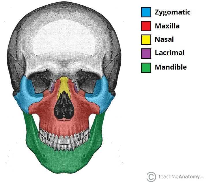
Understanding various elements within cranial structure is essential for grasping their significance in human anatomy. Each section plays a vital role in protection, support, and overall functionality.
- Cranial Vault: Protects brain from external damage.
- Facial Skeleton: Provides structure for face and supports sensory organs.
- Mandible: Facilitates movement for chewing and speaking.
- Orbit: Houses and protects eyes.
- Nasal Cavity: Aids in respiration and olfaction.
Each segment contributes uniquely to overall health and functionality, showcasing an intricate design that supports daily life.
Anatomy of the Cranial Vault
The cranial vault serves as a protective casing for the brain, encompassing vital structures and contributing to overall functionality. Its intricate design allows for both support and flexibility, accommodating various forces and pressures.
Key components include:
- Frontal bone
- Parietal bones
- Occipital bone
- Temporal bones
- Sphenoid bone
These elements work in harmony, forming a robust yet adaptable enclosure. Understanding their relationships and functions is crucial for comprehending head anatomy and related health implications.
Notable features of this region include:
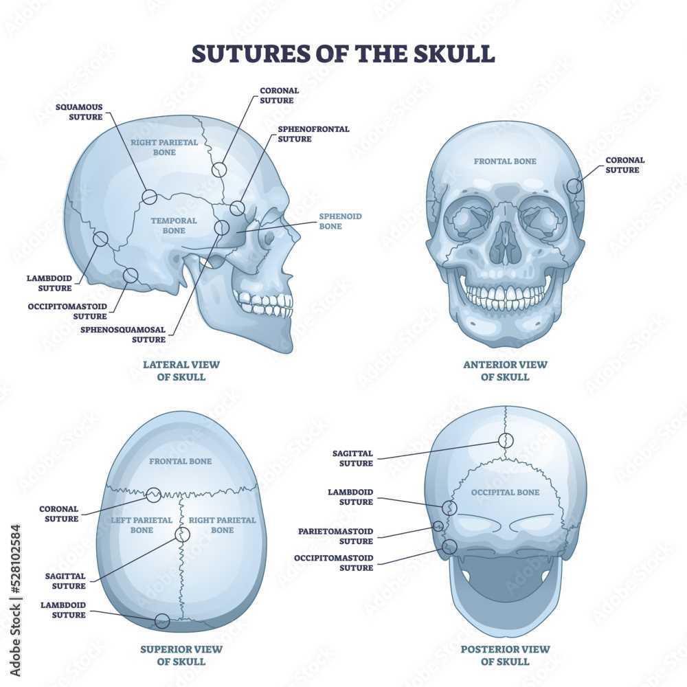
- Cranial sutures: Fibrous joints that unite bones, allowing for slight movement.
- Fossae: Depressions that accommodate brain structures.
- Foramina: Openings that facilitate passage of nerves and blood vessels.
In summary, the cranial vault exemplifies a complex interplay of structures, crucial for safeguarding neural elements while maintaining structural integrity.
Facial Bones and Their Importance
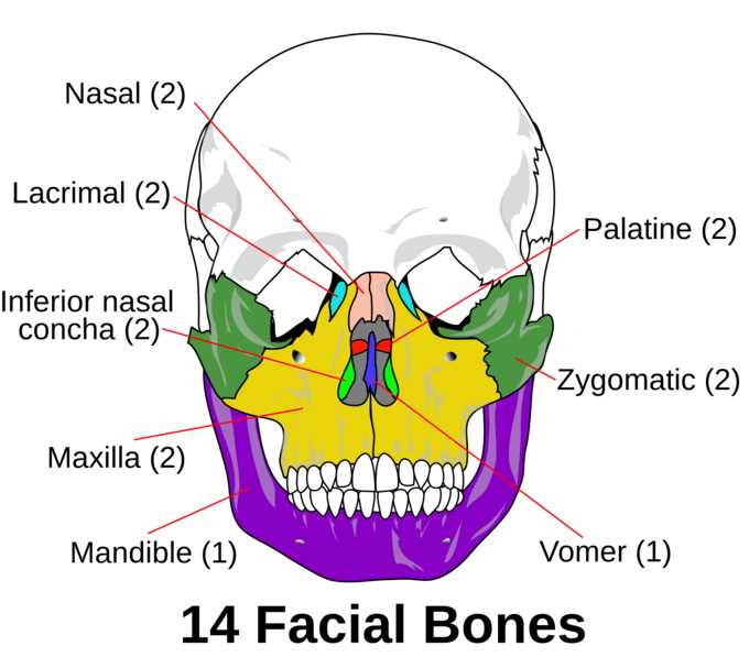
Facial structures play a crucial role in defining individual appearance and functionality. These components provide support for various features and are essential for numerous activities, including eating, speaking, and expressing emotions. Understanding their significance helps in appreciating their intricate design and purpose in human anatomy.
Structural Support and Protection
These components offer vital support to the facial region, ensuring stability and integrity. They form a protective barrier for delicate organs, such as the eyes and nasal passages. Without these structures, the facial region would lack the necessary framework to withstand everyday forces, making them indispensable for overall health and safety.
Role in Communication and Expression

Facial elements are not only functional but also essential for non-verbal communication. They facilitate various expressions, allowing individuals to convey emotions effectively. This aspect enhances social interactions, making these structures vital for human connection and understanding.
Common Skull Fractures Explained
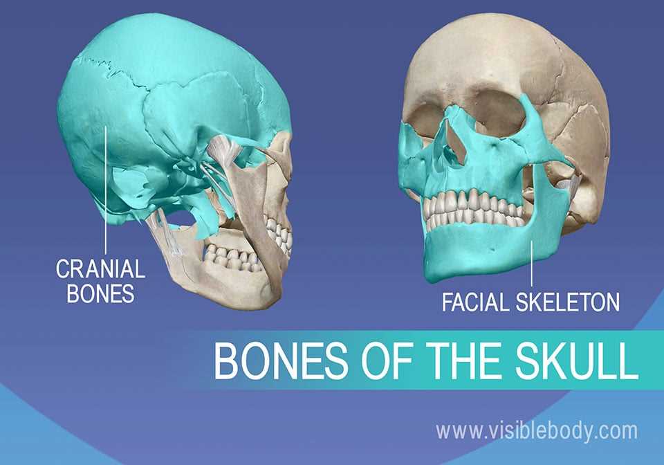
Injuries to cranial structures can vary in severity and type, often leading to significant health concerns. Understanding common fractures helps in recognizing symptoms and determining appropriate responses. This overview highlights typical varieties encountered in medical settings.
Linear Fractures are the most frequent and usually occur due to blunt trauma. These breaks typically run in a straight line and may not require surgical intervention unless complications arise.
Depressed Fractures involve segments of bone being pushed inward, potentially pressing on brain tissue. These injuries often necessitate surgical correction to alleviate pressure and prevent further damage.
Basilar Fractures affect the base of the cranium and can result in serious complications, including cerebrospinal fluid leaks. Symptoms may include bruising around the eyes or behind the ears.
Compound Fractures are characterized by an open wound, exposing inner structures to the external environment. These require immediate medical attention to prevent infections and other complications.
Comparative Anatomy: Human vs. Animal Skulls
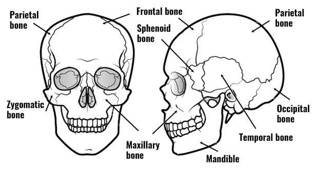
This section explores the fascinating differences and similarities between human cranial structures and those of various animals. Understanding these variations provides insights into evolutionary adaptations and functional requirements across species.
Key aspects of comparison include:
- Size and Shape: Variations in dimensions reflect dietary needs and environmental adaptations.
- Structure: Distinct features such as ridges, cavities, and openings serve different purposes.
- Function: The role of each element varies according to lifestyle, from predation to foraging.
Notable differences:
- Humans possess a relatively large cranial capacity for advanced cognitive functions.
- Predatory animals often have elongated forms for enhanced sensory perception.
- Herbivores display broad structures to support strong chewing mechanisms.
Ultimately, these variations illustrate the remarkable adaptability of organisms within their environments, shedding light on their evolutionary journeys.
Development of the Skull Over Time
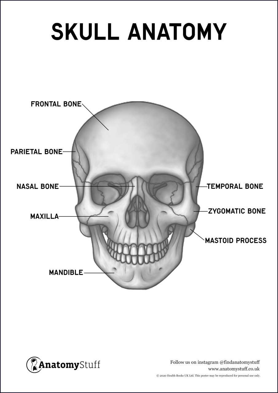
The evolution of cranial structures reflects a fascinating journey through biological history. Over millions of years, these formations have adapted to various environmental demands, functional requirements, and evolutionary pressures.
This process can be observed through several key stages:
- Primitive Origins: Early vertebrates exhibited simple cranial features, primarily serving protective roles.
- Development of Regions: As species diversified, distinct areas emerged, allowing for specialized functions, such as feeding and sensory perception.
- Adaptations to Habitats: Changes in environment prompted variations, enhancing survival through improved mobility and resource acquisition.
- Modern Structures: Present-day formations are the result of complex evolutionary pathways, exhibiting both shared traits and unique adaptations across different lineages.
Understanding these evolutionary transitions provides insight into both past and present biological functions, illustrating how form and function are intricately linked through time.
Skull Anatomy in Forensic Science
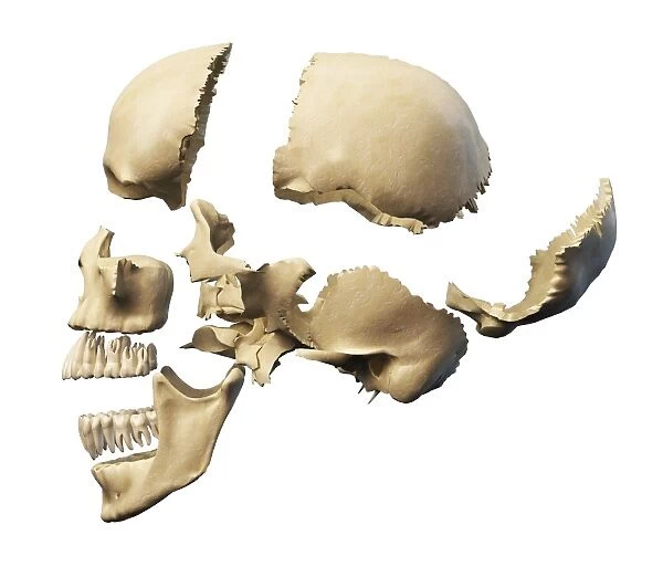
Understanding cranial structure plays a crucial role in forensic investigations, providing insights into identity and cause of death. This knowledge aids experts in reconstructing events surrounding death and can assist in legal contexts.
- Identification: Distinctive features can indicate age, sex, and ancestry.
- Trauma Analysis: Examining fractures and markings helps determine injury patterns.
- Time of Death: Specific changes in cranial remains can suggest post-mortem intervals.
In-depth exploration of these aspects is essential for professionals in forensic anthropology and criminal investigations, ultimately leading to more accurate conclusions.
Visualization Techniques for Skull Anatomy
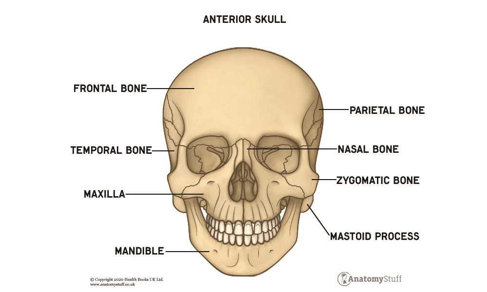
Effective understanding of cranial structure requires various methods to enhance learning and retention. Employing multiple techniques can significantly aid in grasping complex relationships within the cranial region.
- 3D Models: Utilizing three-dimensional representations allows for interactive exploration of bone arrangements.
- Digital Software: Programs designed for anatomical visualization offer detailed views and manipulation of cranial features.
- Anatomical Charts: Color-coded illustrations provide clear depictions of different components and their functions.
Incorporating these methods encourages deeper engagement and a more comprehensive understanding of cranial morphology.
Skull-related Medical Conditions
Various ailments affecting cranial structures can lead to significant health challenges. Understanding these conditions is crucial for diagnosis and treatment, as well as for recognizing symptoms early.
- Fractures: Breaks in cranial bones can result from trauma and may cause complications, including bleeding and nerve damage.
- Congenital Disorders: Some individuals are born with malformations, which can affect appearance and function.
- Infections: Conditions like meningitis involve inflammation of protective membranes and can lead to severe complications if untreated.
- Neurological Disorders: Disorders such as epilepsy can be linked to structural abnormalities in cranial components.
- Tumors: Growths may develop in or around cranial tissues, affecting cognitive and physical functions.
Proper diagnosis typically involves imaging techniques such as X-rays, CT scans, or MRIs, which provide valuable insights into cranial health. Treatment options vary widely based on the specific condition and its severity, ranging from medication to surgical interventions.