Parts of the Heart Diagram Explained
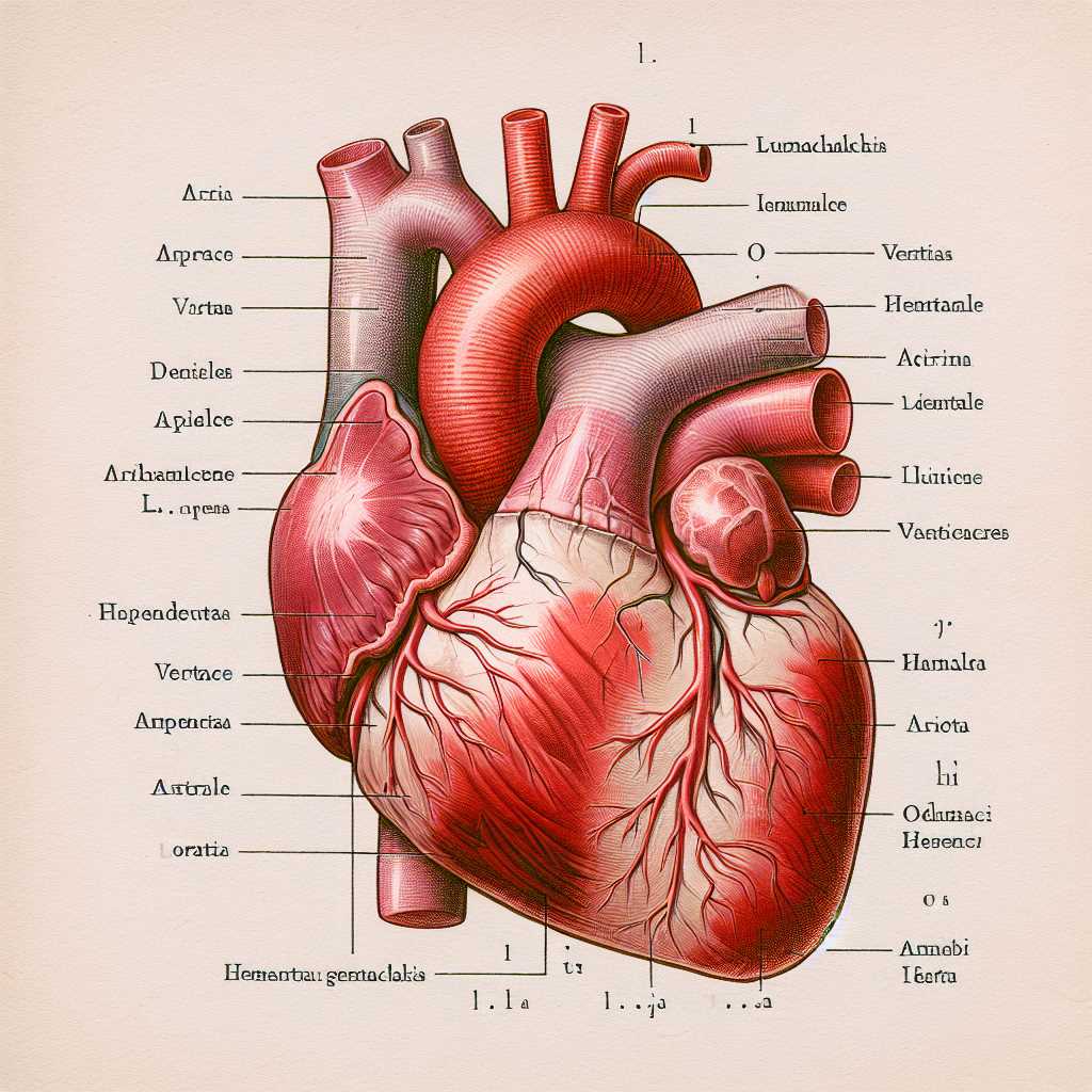
Exploring intricate components of a vital organ reveals fascinating insights into its functionality. This section delves into essential elements responsible for sustaining life through effective blood circulation. Each section plays a unique role, contributing to overall well-being.
Grasping how these crucial structures operate in harmony can enhance appreciation for the complexity of the human body. By examining individual roles and interconnections, one can gain a deeper understanding of how this remarkable system supports various physiological processes.
Moreover, learning about these anatomical features fosters awareness of potential health issues and the importance of maintaining a healthy lifestyle. Awareness can empower individuals to make informed decisions regarding their well-being.
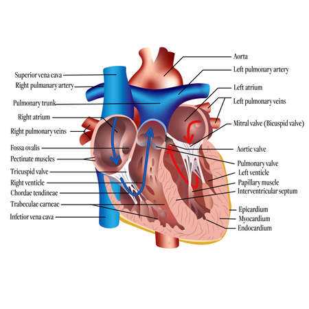
This section aims to provide a comprehensive overview of the essential components that contribute to the functionality of a vital organ in the circulatory system. Understanding these elements is crucial for grasping how they work together to maintain effective blood circulation throughout the body.
Key Components
- Atriums
- Ventricles
- Valves
- Septum
- Blood Vessels
Functions of Major Elements
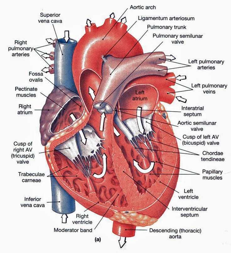
- Atriums: Receive blood returning from the body and lungs.
- Ventricles: Pump blood out to the lungs and the rest of the body.
- Valves: Ensure unidirectional blood flow and prevent backflow.
- Septum: Divides the organ into right and left sides, maintaining separation between oxygenated and deoxygenated blood.
- Blood Vessels: Transport blood to and from the organ, facilitating circulation.
Overview of Heart Functions
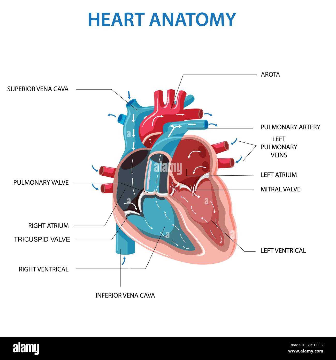
This section explores essential roles of a vital organ responsible for circulating blood throughout the body. It is crucial for sustaining life by delivering oxygen and nutrients to tissues while removing waste products.
Primary Roles
- Regulation of blood flow
- Maintenance of blood pressure
- Support of immune function
Mechanisms of Action
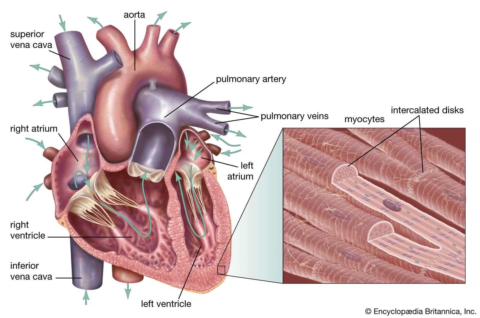
- Contraction and relaxation cycles facilitate rhythmic blood movement.
- Coordination between various chambers ensures efficient circulation.
- Electrical impulses initiate and regulate heartbeat.
Understanding these fundamental functions provides insight into how this organ contributes to overall health and well-being.
Main Chambers of the Heart
This section explores fundamental components crucial for circulation within the body. These regions play vital roles in the movement of blood, ensuring that oxygen and nutrients reach every part effectively.
Upper Sections
Located at the top, these areas receive incoming blood from various sources. They act as initial holding zones, preparing fluid for further distribution to lower sections. Their structure is designed to optimize flow and minimize resistance.
Lower Sections
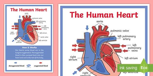
The chambers situated below are responsible for pumping blood outwards. They generate the force needed to propel fluid throughout the body, maintaining circulation and supporting organ function. Their muscular walls enable powerful contractions essential for this process.
Role of Valves in Circulation
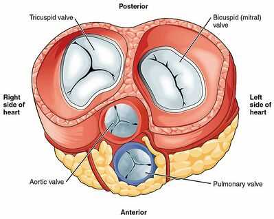
Valves serve as crucial components in the intricate system responsible for transporting blood throughout the body. They ensure unidirectional flow, preventing any backflow and maintaining the efficiency of circulation. This mechanism is vital for sustaining proper oxygenation and nutrient delivery to tissues.
Functionality of Valves
Each valve operates through a simple yet effective mechanism, opening and closing in response to pressure changes. This action regulates blood movement between chambers and vessels, facilitating optimal functioning of the circulatory system.
Impact on Health
Any malfunction or disease affecting valves can significantly disrupt circulation, leading to serious health issues. Maintaining valve integrity is essential for overall cardiovascular wellness, highlighting their ultimate importance in physiological processes.
Coronary Arteries Explained
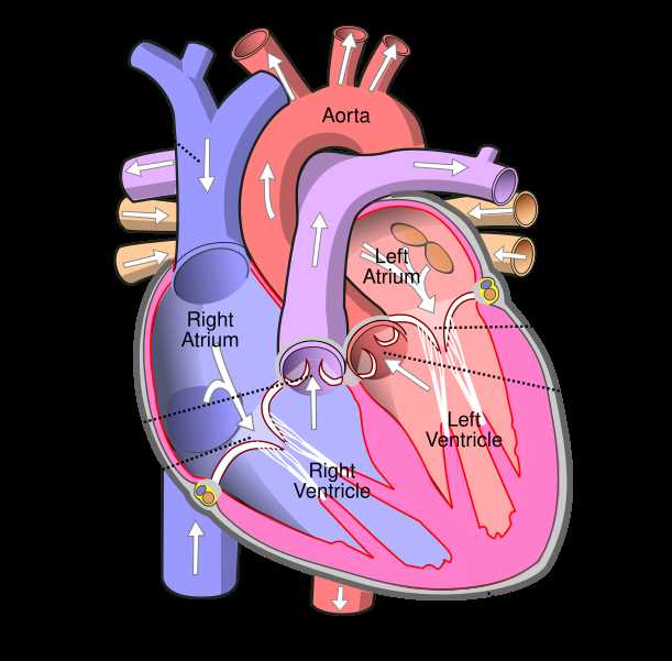
Understanding these vital vessels is essential for grasping how oxygen-rich blood reaches muscle tissue responsible for pumping. Their health directly influences overall wellness and functionality.
- Function: Supply oxygen and nutrients to cardiac muscle.
- Major Arteries:
- Left Main Coronary Artery
- Right Coronary Artery
- Branches:
- Left Anterior Descending Artery
- Left Circumflex Artery
- Marginal Artery
- Importance: Blockages can lead to serious conditions, necessitating awareness and proactive care.
Blood Flow Pathway Through Heart

This section explores the intricate route that oxygen-rich and oxygen-poor fluids traverse within the circulatory structure. Understanding this pathway is essential for grasping how nutrients and gases are exchanged, sustaining life at a cellular level. Each segment of this journey plays a crucial role in maintaining overall physiological balance.
Oxygen-Poor Blood Journey
The journey begins with fluids returning from the body’s tissues, entering through two major veins. Once collected, they flow into a specific chamber where they are temporarily held before moving onward. This region is pivotal in preparing the fluids for the next stage of their journey.
Oxygen-Rich Blood Journey
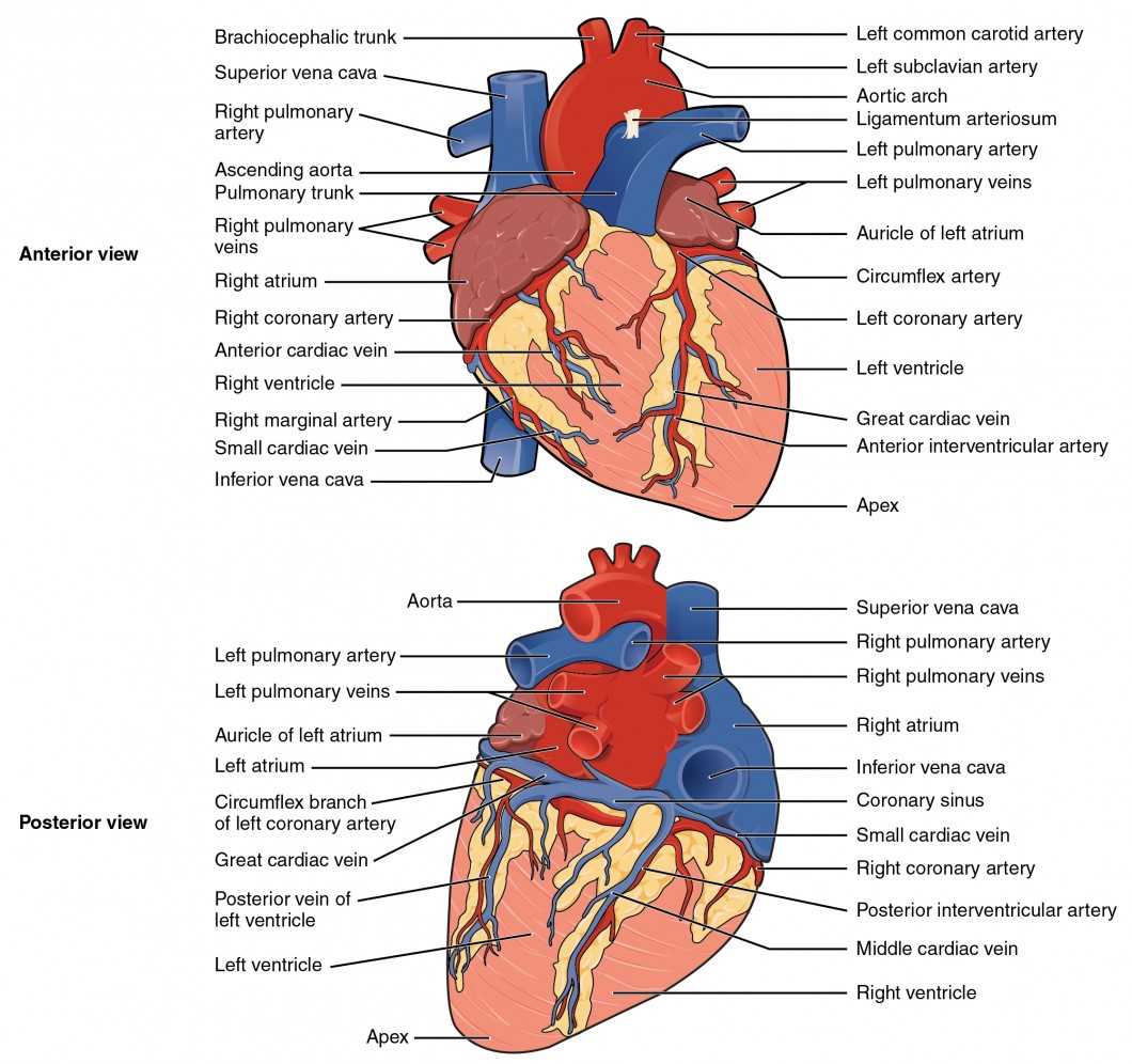
Upon reaching the next chamber, the fluids are pumped into a large artery that distributes them throughout the body. This pathway ensures that all cells receive the vital oxygen and nutrients required for optimal functioning. The cycle continues as the fluids return, completing their circuit.
| Step | Process | Chamber/Structure |
|---|---|---|
| 1 | Oxygen-poor fluids return from tissues | Right Atrium |
| 2 | Pumped into lungs for oxygenation | Right Ventricle |
| 3 | Oxygen-rich fluids return from lungs | Left Atrium |
| 4 | Pumped out to body | Left Ventricle |
Septum’s Importance in Heart Structure
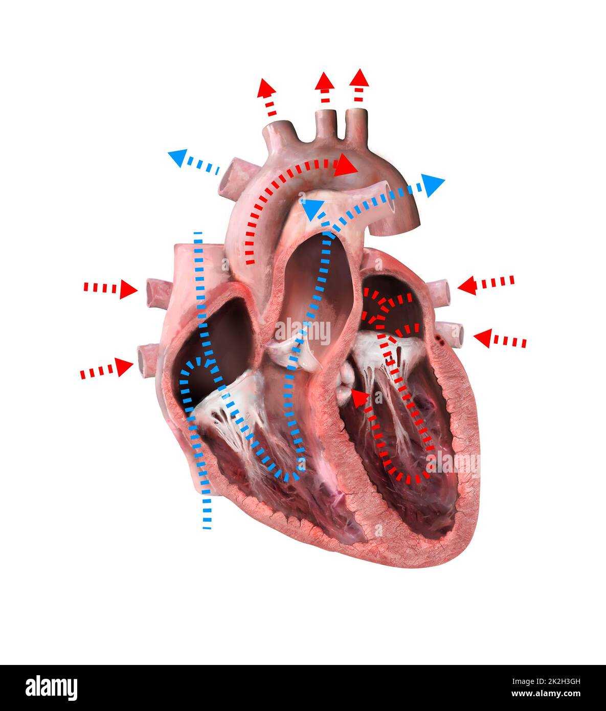
The septum plays a crucial role in maintaining the efficiency of the organ’s function by creating distinct chambers. This separation is vital for the proper circulation of blood, ensuring that oxygen-rich and oxygen-poor blood do not mix. Without this division, overall circulation would be compromised, leading to potential health issues.
Structurally, the septum consists of muscular tissue that provides both strength and flexibility. It enables the organ to pump effectively while adapting to varying pressures during the cardiac cycle. Additionally, its integrity is essential for the proper functioning of valves that regulate blood flow.
In summary, the septum is not just a barrier but a fundamental component that supports overall performance and efficiency of this vital organ. Understanding its significance helps to appreciate the complexity of circulatory mechanisms.
Heart Muscle: Myocardium Details
This segment explores a vital layer responsible for pumping blood throughout the circulatory system. Its unique structure and function are crucial for maintaining an efficient and effective flow, ensuring that all tissues receive necessary nutrients and oxygen.
Characterized by its striated appearance, this muscular layer possesses specialized properties that distinguish it from other types of muscle. Its cells, known as cardiomyocytes, are interconnected by intercalated discs, allowing for synchronized contractions that enable coordinated beats.
| Feature | Description |
|---|---|
| Structure | Striated muscle fibers arranged in a branched network |
| Function | Contracts rhythmically to pump blood |
| Energy Source | Primarily relies on aerobic respiration, utilizing oxygen |
| Regeneration | Limited ability to repair after injury |
| Nervous Control | Influenced by autonomic nervous system, hormones |
Understanding this critical layer’s characteristics and functions provides insight into how vital mechanisms operate within the body, supporting overall health and vitality.
Electrical Conduction System Overview
This section explores the intricate network responsible for regulating rhythmic contractions within the muscular organ. Understanding this system is essential for grasping how signals initiate and coordinate movements effectively.
Components of the Conduction Network
At the core of this system are specialized cells that generate and transmit electrical impulses. These cells ensure efficient communication, allowing synchronized contractions throughout the muscular structure.
Functionality and Importance
Maintaining proper function within this network is vital for overall efficiency. Any disruption can lead to irregularities in contraction, affecting overall performance.
| Component | Function |
|---|---|
| SA Node | Primary pacemaker initiating impulses |
| AV Node | Delays impulses before spreading to ventricles |
| Bundle of His | Transmits impulses to right and left bundle branches |
| Purkinje Fibers | Distributes impulses throughout ventricular walls |
Differences Between Atria and Ventricles
The chambers of this vital organ play distinct roles in the circulation of blood, each contributing to its overall functionality in unique ways. Understanding the characteristics and responsibilities of these two types of chambers reveals important physiological distinctions.
Atria are typically smaller, upper chambers that receive blood returning from the body and lungs. Their primary function is to collect and channel this blood into the lower chambers. In contrast, ventricles are larger, muscular chambers responsible for pumping blood out of the organ. They generate the necessary force to send oxygenated blood to various body parts and deoxygenated blood to the lungs.
Additionally, atria have thinner walls compared to their counterparts, as they only need to facilitate the flow of blood into the ventricles. Conversely, ventricles possess thicker walls, enabling them to withstand higher pressure during contractions. This structural difference is crucial for their respective functions within the circulatory process.
In summary, while both types of chambers are integral to circulation, their size, muscularity, and roles highlight fundamental differences that contribute to the overall efficiency of blood flow throughout the body.
Understanding Heart Surroundings: Pericardium
Encasing a vital organ is a fibrous and serous structure that plays a crucial role in maintaining its functionality. This unique feature provides essential protection, facilitates movement, and ensures that the organ operates efficiently within a confined space. By examining this protective layer, one can appreciate its significance in overall cardiovascular health.
This structure consists of two main layers:
- Fibrous Layer: A tough outer covering that offers stability and prevents over-expansion.
- Serous Layer: A thinner, more delicate membrane that allows smooth movement between the organ and surrounding tissues.
The pericardium serves several important functions:
- Acts as a protective barrier against infections and injuries.
- Reduces friction during heartbeats, enabling seamless contractions.
- Helps maintain appropriate positioning and orientation within the thoracic cavity.
Understanding this structure’s anatomy and function is essential for comprehending how it contributes to overall circulatory health. Any abnormalities or conditions affecting this layer can significantly impact the organ’s performance and overall well-being.
Common Heart Conditions and Diagram
This section explores prevalent ailments affecting a vital organ and their visual representations. Understanding these conditions enhances awareness and promotes informed discussions regarding cardiovascular health.
Common Ailments
Among frequent issues are coronary artery disease, hypertension, and arrhythmias. Each condition impacts overall well-being and can lead to serious complications if left untreated.
Visual Representation
Illustrations serve as essential tools for grasping complex functions and abnormalities. They provide clarity on how various conditions manifest, aiding in education and prevention strategies.
Visual Learning with Heart Diagrams
Utilizing visual representations enhances comprehension of complex biological systems. Engaging with illustrative materials fosters better retention and understanding, making intricate concepts more accessible.
- Improves memory recall
- Facilitates quick recognition of structures
- Encourages active participation in learning
By exploring various visual formats, learners can:
- Identify key components effectively
- Understand functional relationships between elements
- Visualize blood flow and its significance
Ultimately, embracing graphical tools supports a deeper grasp of physiological mechanisms.