Understanding the Essential Parts of a Microscope Diagram
Exploring the intricate world of optical instruments opens up a realm of scientific discovery. Each component plays a vital role in magnifying and clarifying the minutiae of our universe. From basic structures to advanced features, these devices empower researchers and enthusiasts alike.
Familiarity with various elements is essential for anyone interested in observing microscopic phenomena. Each element contributes uniquely to the overall functionality, enhancing the clarity and detail of observations. Whether in a laboratory or educational setting, understanding these components is crucial for effective usage.
In this section, we will delve into the specific features of these instruments, revealing how they work in harmony to facilitate a comprehensive view of tiny structures. Knowledge of these intricacies ultimately enriches the experience of scientific inquiry.
Understanding Microscope Components
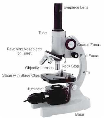
Exploring essential elements of optical instruments reveals their significant roles in magnifying minute details. Each component contributes uniquely to the overall functionality and effectiveness in observation.
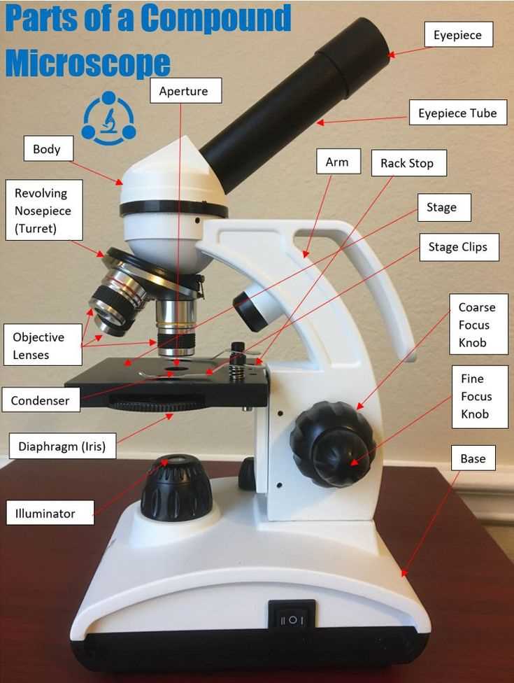
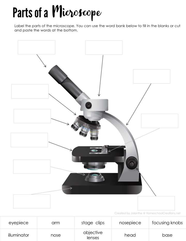
- Base: Provides stability and support.
- Arm: Connects different sections, ensuring ease of use.
- Stage: Holds specimens in place for examination.
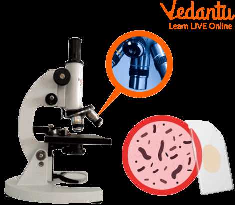
- Objective lenses: Offer varying levels of magnification.
- Eyepiece: Enhances visual experience by further enlarging images.
Familiarity with these elements enhances comprehension of their interplay during usage. Understanding their functions ultimately leads to more effective and precise observation techniques.
Basic Structure of a Microscope
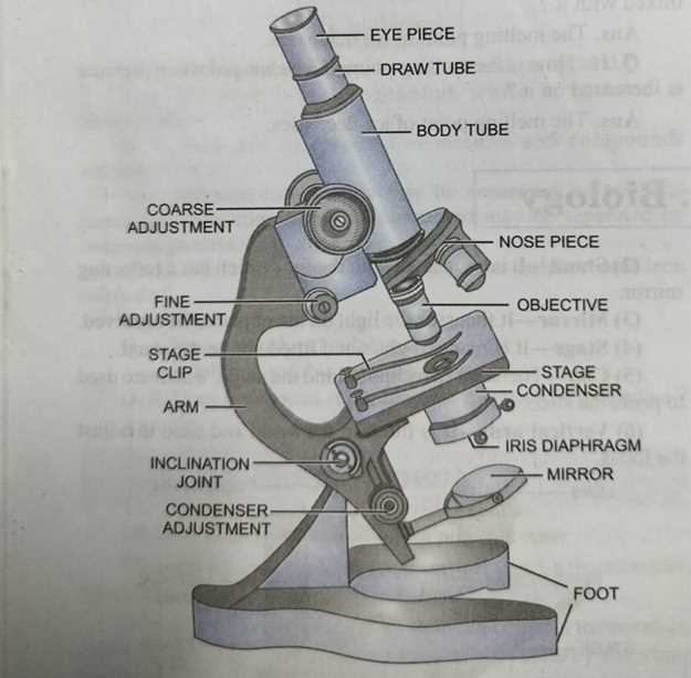
This section explores fundamental elements that comprise an optical instrument designed for magnifying small objects. Understanding these components is essential for effective use and appreciation of this scientific tool.
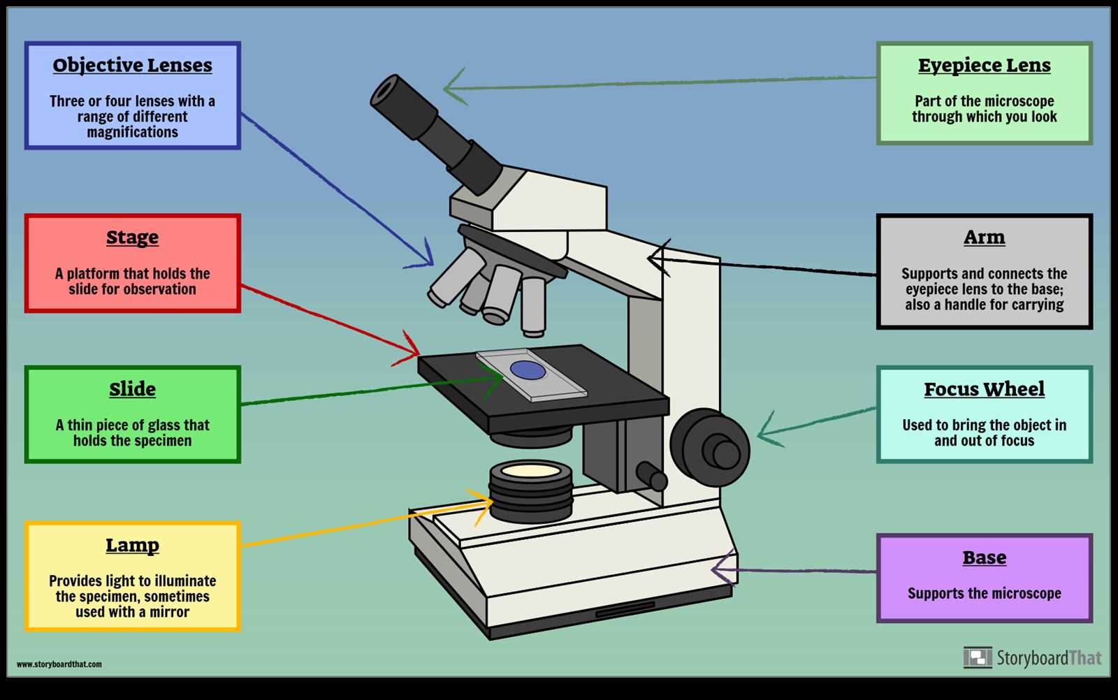
- Base: Provides stability and support, ensuring a solid foundation.
- Arm: Connects the base with optical systems, allowing easy handling and maneuverability.
- Stage: Flat platform where specimens are placed for observation.
- Optical Tube: Houses lenses that magnify images, directing light toward the viewer’s eye.
- Objective Lenses: Various lenses with different magnification powers, typically rotating to adjust focus.
- Eyepiece: The lens at the top, through which the user looks to view the magnified image.
- Iris Diaphragm: Controls light intensity reaching the specimen, enhancing visibility.
- Light Source: Illuminates the specimen, essential for clear observation.
Each component plays a crucial role in enhancing visibility, allowing researchers to delve deeper into microscopic worlds.
Importance of the Objective Lenses
Objective lenses serve a crucial role in enhancing clarity and detail of specimens under observation. Their design and specifications greatly influence overall image quality and resolution, allowing for an in-depth examination of various materials.
These lenses come in different magnifications, each tailored for specific applications. High-quality objectives can reveal intricate structures that might otherwise remain unseen. Effective utilization of these lenses is essential for achieving the ultimate understanding of complex biological and material characteristics.
In research and educational settings, choosing the appropriate objective is vital for accurate results. The ability to switch between different magnifications enables users to delve deeper into analyses, ensuring comprehensive insights.
Function of the Eyepiece Lens
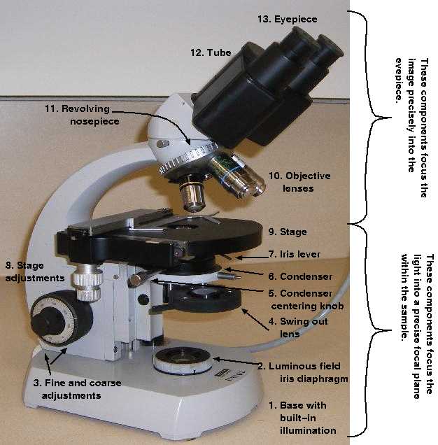
The eyepiece serves as a crucial element for visualizing specimens, allowing users to enhance their observational experience. This optical component magnifies images produced by objective lenses, facilitating a clearer view of intricate details.
Magnification and Clarity
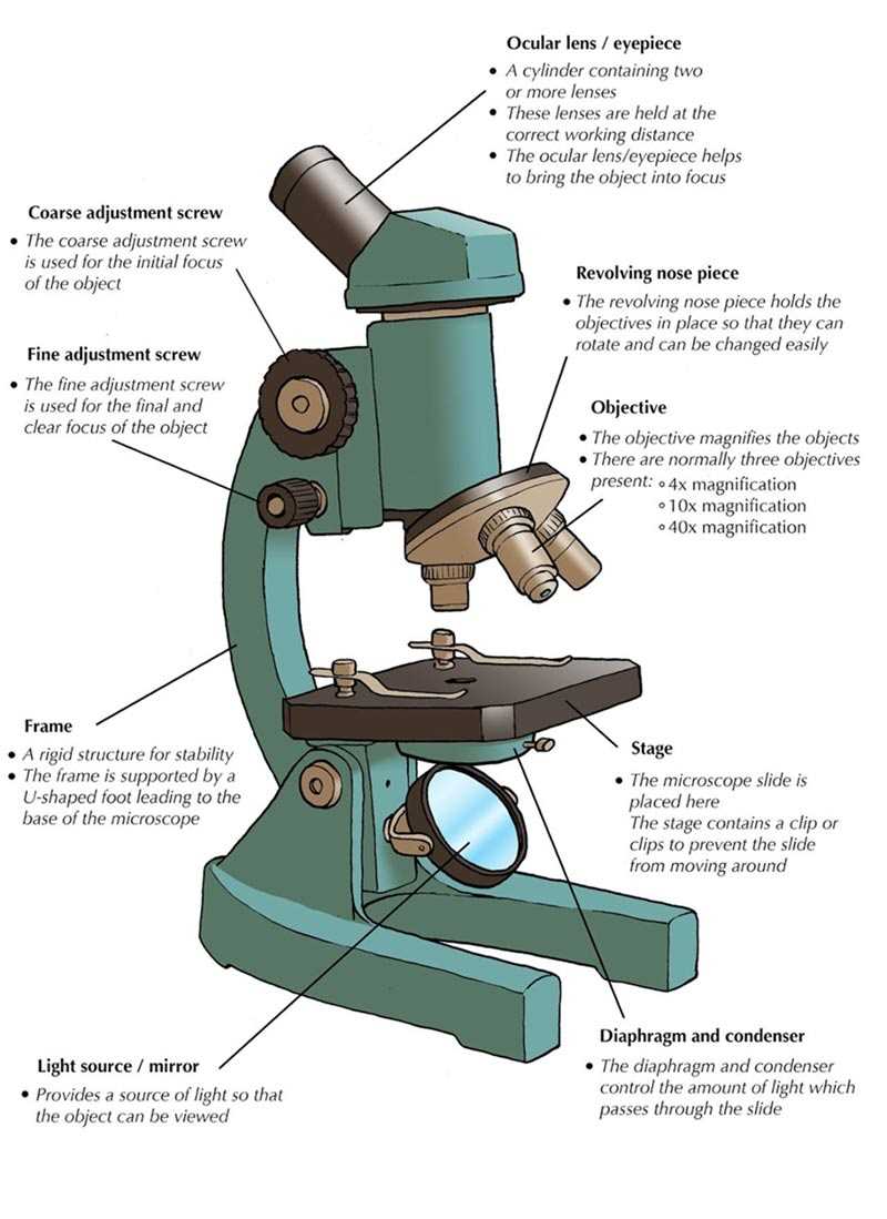
Primarily, eyepieces contribute to overall magnification. They are designed to provide additional enlargement, making it possible to observe fine structures with remarkable clarity. This function is essential for accurate analysis and research.
Field of View
Moreover, this lens influences the field of view, allowing users to see a broader area of the sample. A wider field aids in context comprehension, making it easier to identify features and anomalies within the observed subject.
The Role of the Stage
A crucial component within optical instruments is designed to support specimens during examination. This area facilitates precise placement and stability, allowing users to focus on the intricate details of samples. Its functionality enhances observation, making it essential for effective study and analysis.
Functions and Importance
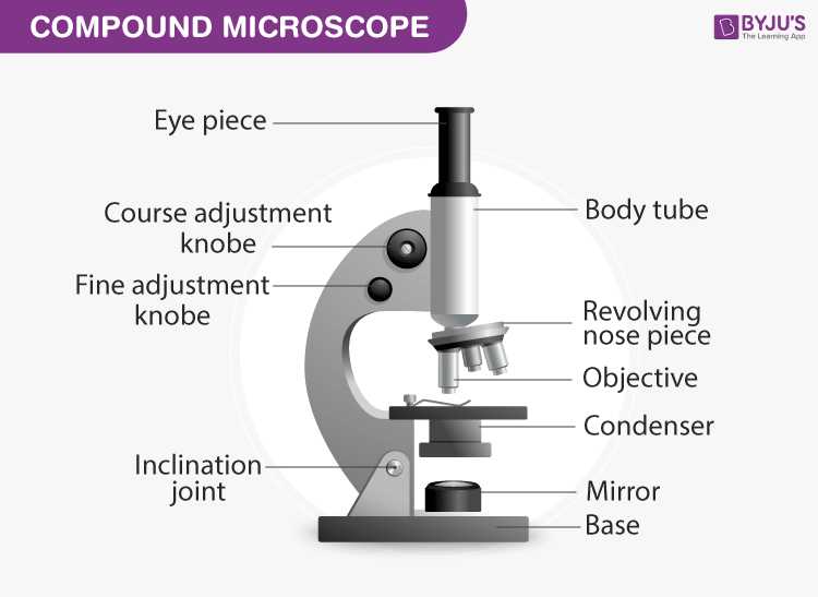
This supporting structure serves multiple purposes, such as providing a stable base and ensuring that light passes effectively through specimens. Its adjustable features allow for positioning adjustments, optimizing clarity and detail during observations.
Specifications
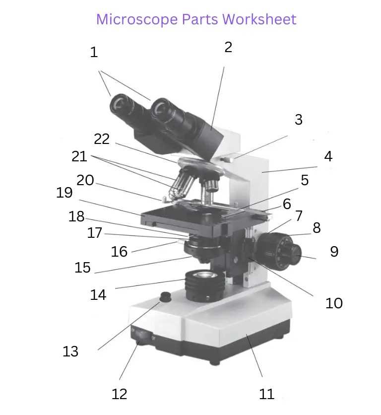
| Feature | Description |
|---|---|
| Material | Often made of durable materials for longevity |
| Size | Varies to accommodate different specimen types |
| Adjustment Mechanism | Allows for vertical and horizontal movement |
| Light Source Compatibility | Designed to align with various illumination systems |
Illumination Systems Explained
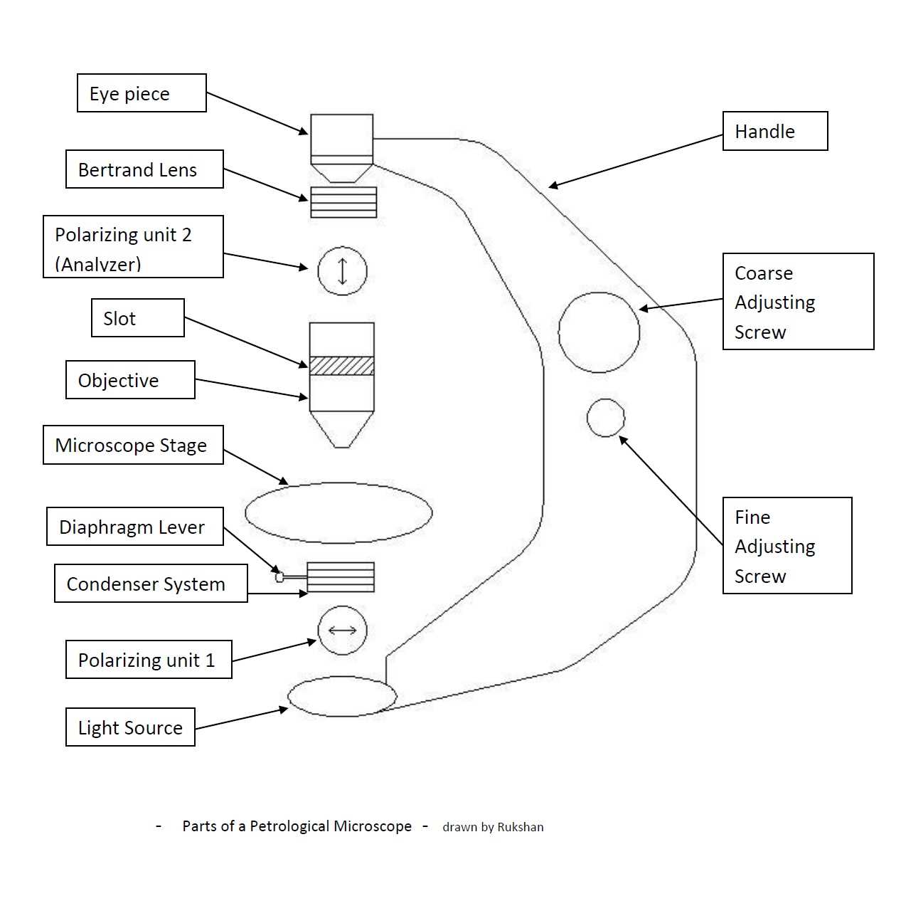
Effective lighting is crucial for optimal observation in scientific instruments. Various techniques and components play a vital role in ensuring clarity and detail when examining specimens. Understanding these illumination methods enhances appreciation of their significance in research and educational settings.
- Transmitted Light: This approach involves shining light through a sample, making it visible by highlighting contrasts between different structures.
- Reflected Light: Here, illumination is directed onto a specimen’s surface, allowing for examination of opaque materials or textured surfaces.
- Fluorescence: This technique uses specific wavelengths to excite fluorescent dyes, revealing particular components within a sample.
- Dark Field: A method that enhances visibility of specimens by scattering light, creating a contrast against a dark background.
Each illumination technique offers unique advantages, catering to various observation requirements and specimen types. By utilizing the appropriate system, researchers can achieve more precise and informative results.
Mechanics of the Focusing Knobs
Focusing knobs play a crucial role in enhancing clarity and detail during observation. Their design allows for precise adjustments, ensuring that specimens are viewed with the utmost sharpness. Understanding the underlying mechanics reveals how these controls facilitate optimal image quality.
Operation Principles
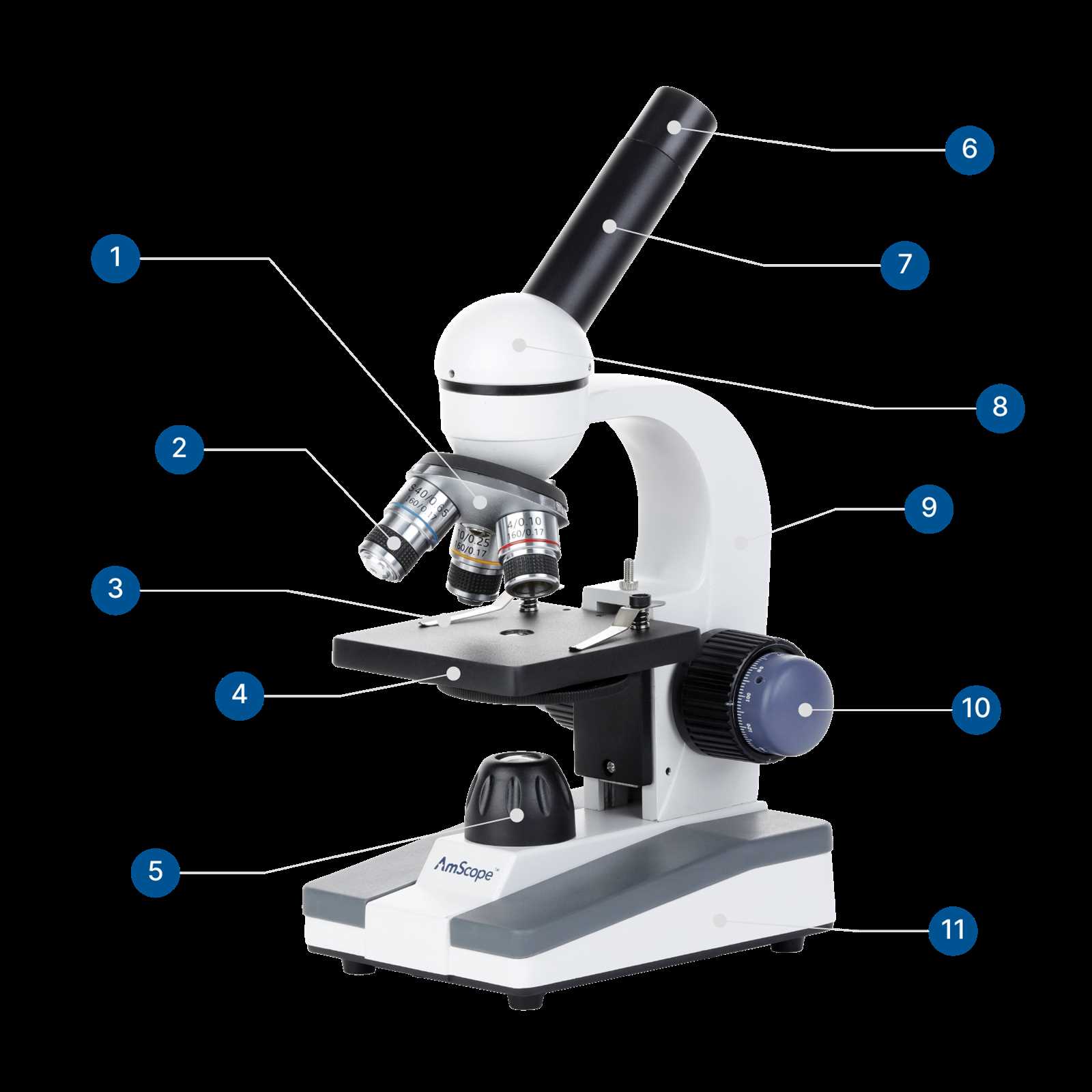
Typically, these controls utilize a system of gears and levers, translating rotational movement into linear adjustments. This mechanism enables gradual or rapid changes in focal distance, catering to varying needs of users.
Importance of Calibration
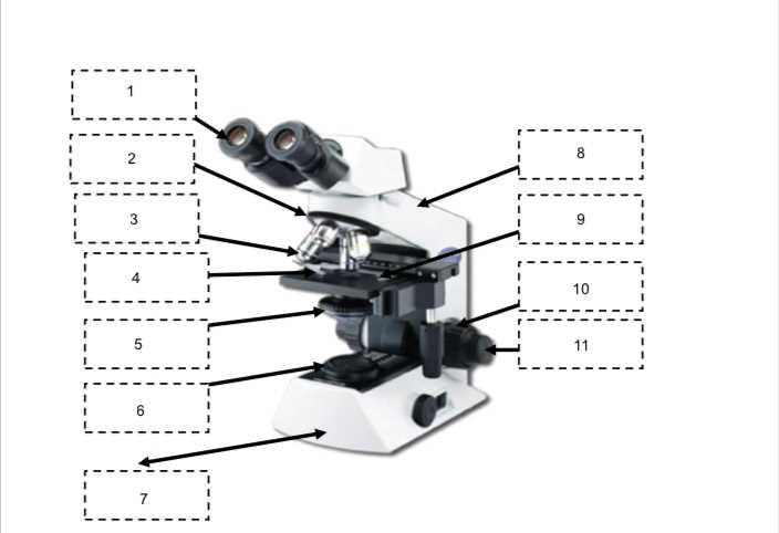
Accurate calibration of these knobs is essential for achieving the best possible results. Proper alignment ensures that each turn directly correlates with the desired visual outcome, minimizing frustration and maximizing efficiency during examination.
Types of Microscopes Overview
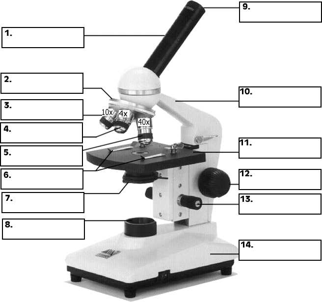
Exploration of various optical instruments reveals a fascinating array of designs, each tailored for specific purposes and applications. These tools enable researchers to observe structures at magnifications beyond the capabilities of the naked eye, enhancing our understanding of biological and material sciences.
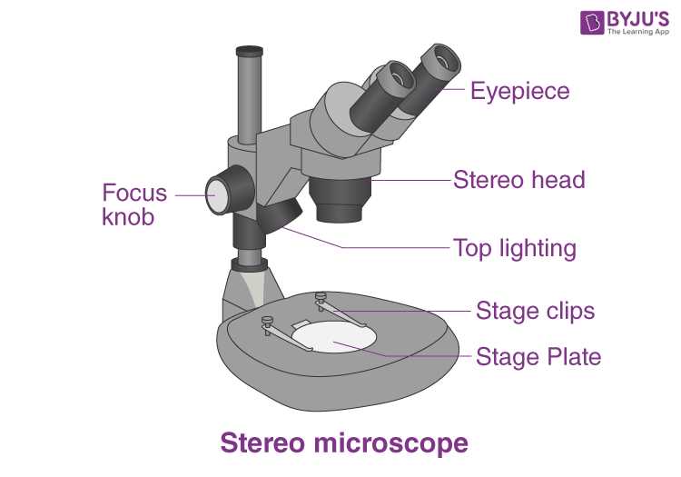
Optical instruments utilize visible light to illuminate specimens, allowing users to view samples through lenses. Common types include compound and stereo variations, each offering unique benefits depending on the level of detail required.
Electron devices employ beams of electrons instead of light, achieving significantly higher magnifications. This category includes transmission and scanning models, ideal for examining fine details at a molecular level.
Additionally, digital tools have emerged, integrating cameras and software to capture and analyze images. This modern approach enhances collaboration and facilitates sharing of findings across various fields.
Ultimately, selection of an appropriate instrument hinges on intended use, specimen type, and desired resolution, paving the way for significant discoveries in science and industry.
Key Features of the Arm
The arm serves as a vital support structure, providing stability and connectivity between various components of an optical instrument. Its design ensures ease of use while facilitating proper alignment during observation. Understanding its unique characteristics enhances overall functionality and user experience.
Structural Elements
- Sturdy Design: Constructed to withstand handling, ensuring longevity.
- Ergonomic Shape: Designed for comfortable grip, promoting user-friendly operation.
- Attachment Points: Features connectors for additional accessories or lenses.
Functional Aspects
- Stability: Keeps optical elements securely in place during use.
- Alignment: Aids in proper positioning of lenses and other optical components.
- Mobility: Allows for easy adjustment and repositioning without compromising integrity.
Significance of the Base
The foundation of any optical instrument plays a crucial role in ensuring stability and support during observation. Its design influences overall functionality, affecting user experience and efficiency in conducting experiments.
This structural component not only provides a steady base but also serves as a platform for integrating various elements of the apparatus. By maintaining equilibrium, it allows for precise adjustments and facilitates seamless operation, which is essential for obtaining accurate results.
Moreover, a well-designed foundation contributes to the longevity of the instrument, protecting sensitive components from external disturbances. In essence, this element is vital for effective use and reliability in diverse scientific applications.
Differences Between Monocular and Binocular
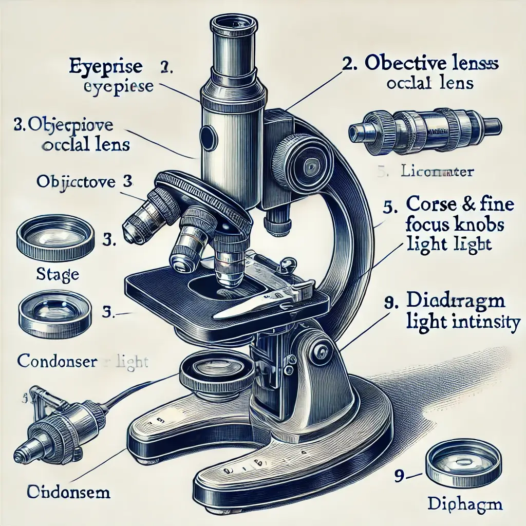
Understanding variations in optical devices enhances appreciation for their functionality and application. Two common types are distinguished by their design and user experience, influencing how users engage with visual observations.
| Feature | Monocular | Binocular |
|---|---|---|
| Vision | Single-eye observation | Two-eye observation |
| Depth Perception | Limited | Enhanced |
| Weight | Lighter | Generally heavier |
| Cost | More affordable | Typically more expensive |
| Usage | Portable, suitable for casual use | Preferred for detailed viewing, such as in labs |
Each type serves unique purposes, making choice dependent on specific needs and contexts. Awareness of these distinctions can guide optimal selections for varied observational tasks.
Understanding the Condenser Lens
A crucial element in optical systems, this component plays a vital role in enhancing illumination and clarity. By concentrating light onto specimens, it facilitates detailed observations, making it indispensable for researchers and students alike.
Functionality and Importance
The primary function of this lens is to collect and direct light towards the object being examined. Proper alignment significantly impacts image quality, ensuring that details are sharply defined. Without this focus, even the most intricate samples can appear blurred and indistinct.
Adjustability and Control
Many devices feature adjustable elements that allow users to modify light intensity and focus. This adaptability enables users to delve deeper into various samples, optimizing visibility for different types of analyses. Mastering this component ultimately leads to more accurate observations and insights.
Using the Diaphragm for Light Control
Effective illumination is crucial for enhancing visibility and detail in observations. This component plays a vital role in regulating brightness and contrast, allowing for optimal clarity when examining specimens. By adjusting light intensity, users can significantly improve image quality, facilitating a deeper understanding of the subject matter.
Adjusting Light Intensity
By manipulating this feature, one can fine-tune the amount of light passing through, which is essential for various types of samples. A lower setting may be beneficial for transparent or delicate materials, while increased brightness can reveal intricate structures in opaque specimens. Balancing these adjustments ensures that details remain visible without overwhelming the viewer.
Enhancing Contrast
In addition to brightness, this element aids in enhancing contrast. Optimal settings allow for clearer differentiation between various features within a sample, making it easier to analyze specific aspects. Understanding how to effectively use this tool is fundamental for achieving the best observational results.